X-rays
X-rays
The invisible roentgen rays named after Wilhelm Conrad Röntgen (German physicist, 1st physics Nobel prize 1901 (initially referred to as "X-rays" in German, still called X-rays today in English]) are used for therapeutic, but mainly for diagnostic purposes in medicine, dentistry, structure and material testing.
The electromagnetic (ionising, i.e. uncharged molecules dispersed in ions and electrons) roentgen rays have shorter wavelengths than visible light. They penetrate body tissue and are biologically harmful, e.g. carcinogenic, (cancer-causing), mutagenic (gene-changing) and teratogenic (embryo-damaging). This is why the frequency (i.e. the number of X-ray images) and the respective single dose must be limited to the absolutely necessary in order to minimise radiation exposure. Areas that should not be irradiated must be shielded using suitable materials (mainly lead, e.g. in X-ray shields, radiography aprons, X-ray protective doors etc.).
Diffraction of X-rays allows conclusions to be made about the structure of molecules (DNA), crystals and materials. Emission of element-typical spectra of X-rays after irradiation with electrons or X-rays (X-ray fluorescence) enables analysis of the chemical elements contained in the substances.
Orthopaedic pain levels ("X-ray inflammation irradiation") and tumour cells, which are sensitive to radiation, can be combated using X-rays. The (e.g. intraoperative) X-raying of body structures (using radiographic image intensifiers) is used for analysis of processes and optimisation (e.g. reposition of fractures).
Occasions for using diagnostic X-rays can be acute complaints, traumata or disease status, but also exclusion and early diagnosis of damage (bitewing X-ray for early detection of caries) as well as planning and follow-up of dental treatment measures (surgery, endodontology, implantology, orthodontics, prosthetics).
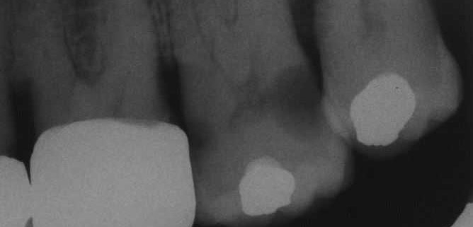 Extensive carious cavity in tooth 27
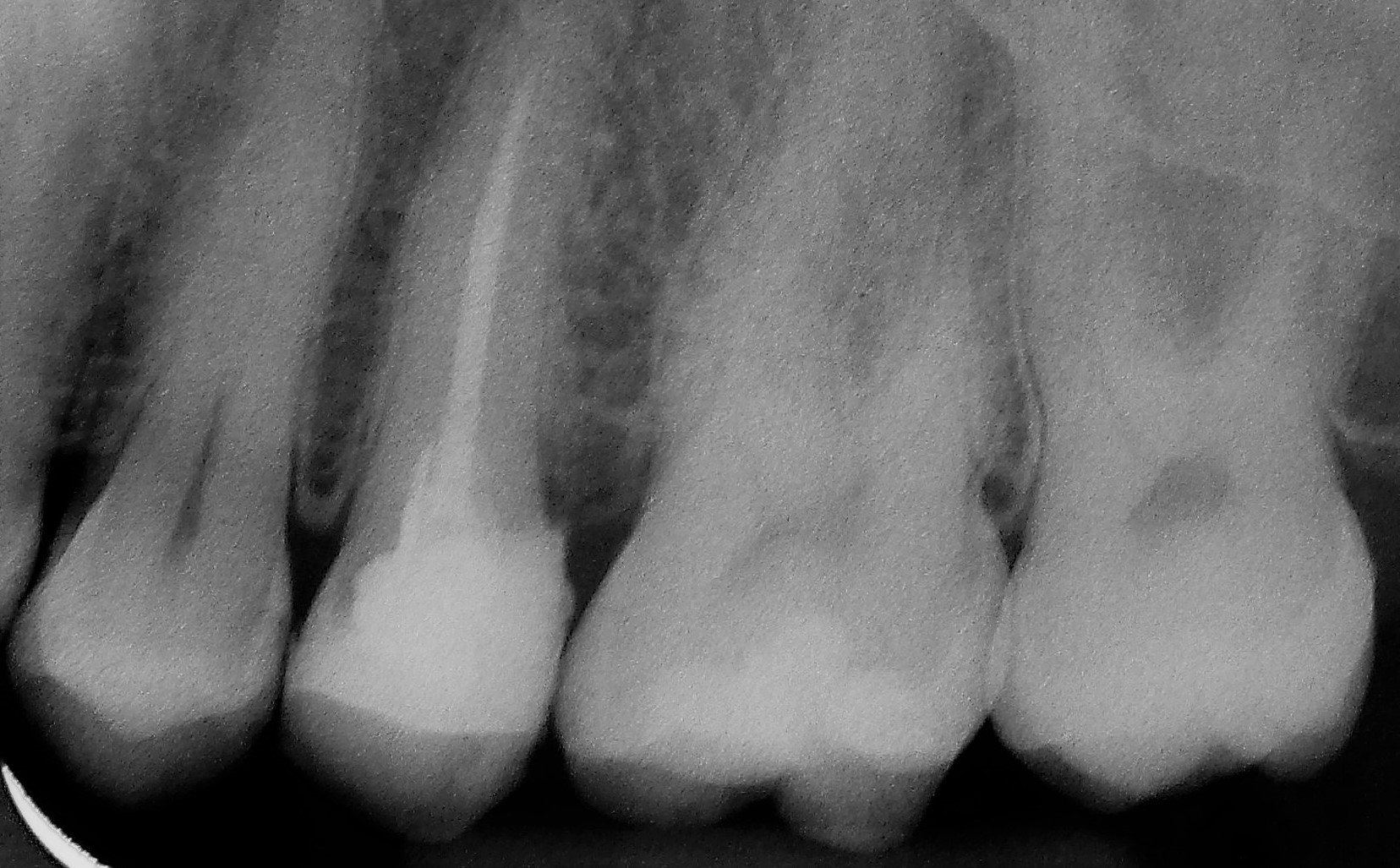 X-ray image
In dentistry intraoral X-ray tubes (X-ray sources) are rarely used, though extraoral X-ray tubes are mainly used to irradiate analogue, light-sensitive X-ray films (chemical development and fixation in the darkroom), X-ray imaging plates (stimulation of fluorescent substance, scanning using lasers) or digital X-ray sensors (CCD or CMOS semiconductor devices). The X-ray voltage of "conventionally" used X-rays is 60 to 70 kilovolts. Two-dimensional images are created using static systems (dental film, lateral cephalometric radiograph, temporomandibular images) and orthopantomograms and three-dimensional images (CT = computer tomography, CBCT = cone-beam computed tomography) are created using rotary systems. The images are viewed on X-ray viewers (analogue) or (if necessary, combined with image processing) on the computer monitor (digital).
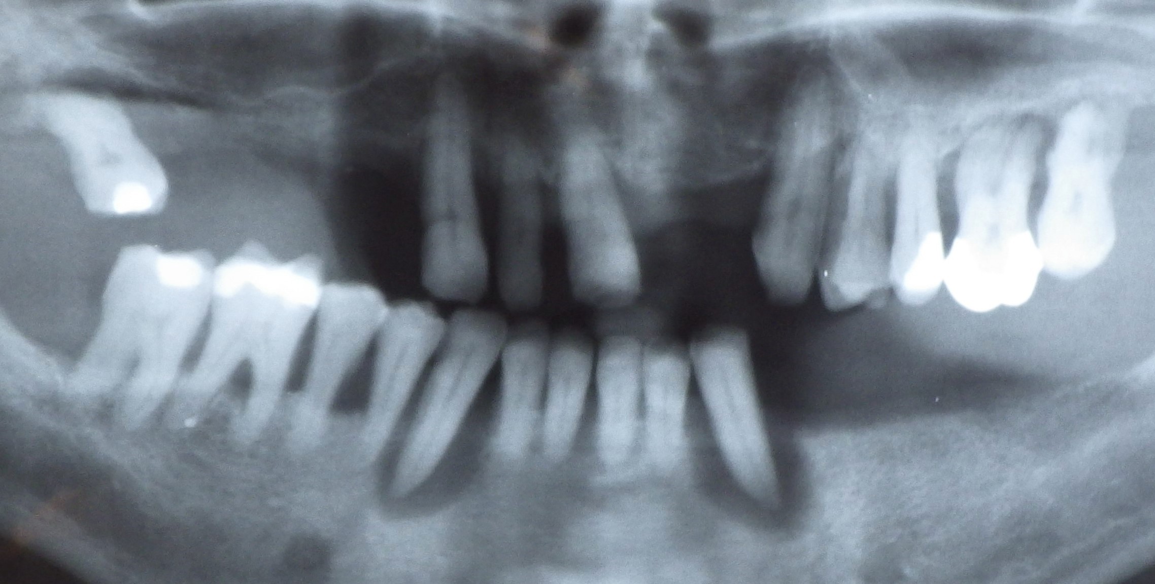 Panoramic X-ray, orthopantogram
.jpg) Orthopantogram of an 8-year-old (deciduous dentition)
Orthopantogram of an 8-year-old (deciduous dentition)
Different body tissues contain varying amounts of chemical elements whose proton count in the atom nucleus differ and consequently their absorption of the X-rays, which shows as image contrast, e.g. between hard structure with a different calcium content (bone, dentine, dental enamel) and soft tissue. A greyscale image is created with a resolution of up to 20 line pairs per millimetre or 25 µm. The usual negative representation leads to correspondingly "reverse" terminology, lighter areas are defined as shadowing and darker areas as brightening.
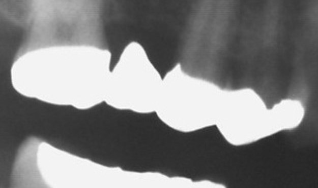 Bridgework spanning teeth 15 - 17
Metal foreign bodies (amalgam, cast restorations) and ceramics placed in the oral cavity normally have a high degree of radiopacity and appear white or light grey. Filling materials (temporary sealers, composite and gutta percha) medication (calcium hydroxide paste) or acrylics and denture teeth for X-ray templates can be made radiopaque using positive X-ray contrast materials.
PROBIEREN SIE ES EINFACH AUS !!!
Von uns erhalten Sie professionelle Unterstützung.
Treten Sie mit uns in Kontakt oder nutzen Sie unser Kontaktformular.
Wort des Tages
| Deutsch | Englisch |
|---|---|
| Alveolarkamm-Abhang | ridge slope |
Schwerpunkttext des Monats
Implantat-Suprakonstruktionen Implantat-Suprakonstruktionen
Eine S. kann rein implantatgetragen sein oder sich sowohl auf Zähnen, als auch auf Implantaten abstützen. Insbesondere bei Brücken spricht man dann von Hybrid- oder Verbund-Zahnersatz.
Bei zementierten S. ist zwischen provisorischer (temporärer), definitiver (permanenter) und semi-permanenter Zementierung zu unterscheiden. Letztere soll eine sichere Befestigung und gleichzeitig das Abnehmen der S. durch den Zahnarzt im Bedarfsfall ermöglichen. Damit handelt es sich um eine sogenannte bedingt abnehmbare (für den Patienten also festsitzende) S. Dazu gehören auch die verschraubten S. Die beiden Befestigungsarten bieten Vor- und Nachteile: Verschraubungen bedingen Spalträume, die bakteriell besiedelt werden können, zur Vorbeugung dagegen werden spezielle Gele zum Einbringen in den Implantat-Innenraum angeboten, die langfristig wirksam bleiben sollen. Erfolgt eine Fixation von S. mit Schrauben, können bei diesen auch Misserfolge durch Lockerung, Überlastung und Bruch auftreten. Da Implantate keine Eigenbeweglichkeit aufweisen und starr im Kieferknochen verankert sind, wird stets ein spannungsfreier Sitz von S. angestrebt. Er kann bei verschraubten S. auf mindestens zwei Pfeilern mit dem Sheffield-Test (spaltfreier Sitz bei Anziehen einer beliebigen Einzelschraube) überprüft werden. Um spannungsfreie Gerüste herzustellen, werden Verfahren zur intraoralen Verbindung (etwa Verkleben) von Teilen der S. und/oder zur digitalen Fertigung (z.B. Fräsen, Sintern) angewendet. Um unzugängliche Zementüberschüsse, die zu Periimplantitis und Implantatverlust führen können, zu vermeiden, sollte der Restaurationsrand zementierter S. stets im Bereich des Zahnfleischrands enden. Dies lässt sich – vor allem bei Implantatplattformen auf Knochenniveau ("bone level") mit entsprechenden (ggf. individuell angefertigten) Abutments erreichen. Abutments dienen als Verbindung zwischen Implantaten und S. Bei S. auf mehreren Pfeilern ermöglichen abgewinkelte Formen die Parallelisierung hin zu einer gemeinsamen Einschubrichtung. Abutments können entweder die Form eines präparierten Zahnstumpfs nachahmen oder eine Komponente eines Verbindungselements (z.B. Druckknopfsysteme, Kugelköpfe, Stege, Magnete) beinhalten. Die S. umfasst in diesen Fällen die entsprechenden ergänzenden Komponenten. |
 Ober- und Unterkiefertotalprothese (Unterseite, mit Kugelkopfmatrizen)
 Kugelkopf
 Metallkeramikkronen auf Implantaten